VERC Advanced Diagnostics

In-house Digital Imaging and Diagnostic Laboratory

Getting a complete picture of your pet’s medical condition is crucial to providing an accurate diagnosis and effective treatment plan. From our in-house veterinary reference laboratory to multiple digital imaging modalities, we have the advanced diagnostic equipment and the staff to provide thorough and accurate diagnostics for your animal.

Full In-House Laboratory/Vet Reference Lab

In most veterinarian offices, doctors have to decide between the speed of performing a lab test in-house or the reliable accuracy of sending a sample to a vet reference lab. At VERC, we have an on-site certified vet reference lab, with testing performed by experienced personnel on high-end professional equipment.

Our vet reference lab provides quick and accurate results for evaluating blood, urine, CSF and other fluids to screen for medical conditions and diseases. Rather than waiting days for the results of many tests, the Veterinary Emergency Referral Center has the information we need to provide care for your pet with same-day results from tests performed by our in-house veterinary reference lab.

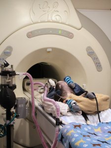
MRI Scans

Magnetic resonance imaging, or MRI, is a valuable advanced diagnostics tool for capturing highly detailed digital images of your pet. The Veterinary Emergency Referral Center has the only veterinary MRI machine in Northwest Florida for capturing images used in diagnosing and treating trauma and disease. Our MRI machine is used to perform in-house scans quickly to assess and diagnose your pet.
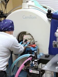
MRIs are especially helpful in identifying the causes of neurological issues involving the brain and spinal cord. Our doctors also use the results of these scans to observe fine details of internal organs, bones, muscles and other soft tissues.


Common Pet Conditions Requiring an MRI

  • Tumors
  • Inflammation
  • Herniated discs
  • Spinal cord disease/stenosis
  • Seizures
  • Difficulty walking
  • Paralysis/paresis
  • Neurological conditions
  • Fainting/syncope

CT Scans and Myelograms

A CT, which stands for computed tomography, is a type of imaging scan where the device takes multiple thin digital X-rays of an area of your pet’s body. Computer software then combines these “slices” to form a three-dimensional image of the area. This allows our veterinarians to visually examine internal structures in a safe, non-invasive way.

The Veterinary Emergency Referral Center also performs myelograms, a special type of CT scan where the veterinarian injects contrast into the spinal canal to identify damage or compression of the spine.


Common Pet Conditions Requiring CT Scans

  • Brain tumors
  • Neurological conditions
  • Inner ear problems
  • Fractures
  • Joint problems
  • Soft tissue injuries
  • Arthritis

Endoscopy and Fluoroscopy

In order to evaluate the organs of the digestive and urinary systems, the Veterinary Emergency Referral Center often performs an endoscopy. Typically, this is done by inserting a thin probe with a camera into your pet. This allows our specialists to see internal structures and collect samples or biopsies.

Fluoroscopy is an advanced diagnostics technique that uses a continuous series of X-ray images to observe motion inside the body of your pets. It is often used to observe the lungs, trachea, stomach, intestines, heart valves and major blood vessels. In certain cases, fluoroscopy is also used to help guide an endoscope to the heart and major vessels or for internal stent placement.


Common Pet Conditions Requiring an Endoscopy

  • Vomiting
  • Diarrhea
  • Unexplained weight loss
  • Difficulty urinating or defecating
  • Difficulty breathing/respiratory distress
  • Difficulty swallowing
  • Choking/coughing

EKG and Echocardiogram

An electrocardiogram, often referred to as an EKG or ECG, is a simple test that records the electrical activity of your pet’s heart. The measurements are taken through electrodes placed on the skin and is not painful or invasive. Our veterinarians use an EKG to evaluate the rhythm of your pet’s heart and identify any conditions such as an irregular heartbeat or enlargement of the heart.

When our vets suspect an animal is suffering from cardiac issues, we may perform an echocardiogram, which uses an ultrasound to view how your pet’s heart is functioning using sound waves. With these two tests, our veterinarians can get a complete picture of your pet’s cardiac health. The Veterinary Emergency Referral Center uses the same high-end equipment used on people in cardiologist’s offices to provide accurate diagnoses and effective treatment plans.


Common Pet Conditions Requiring an EKG

  • Congestive heart failure (CHF)
  • Difficulty breathing/respiratory distress
  • Fainting/syncope
  • Weakness/exercise intolerance
  • Heart murmur
  • Irregular heart beat
  • Enlarged heart/cardiomegaly

Ultrasounds

Ultrasound imaging for pets at the Veterinary Emergency Referral CenterThe Veterinary Emergency Referral Center uses ultrasound imaging, also called sonography or ultrasound scanning, to perform advanced diagnostics where we observe organs and processes inside your pet’s body. Using high-frequency sound waves, ultrasound scans produce visual images of internal organs in real time. This allows our veterinarians to observe active movement, such as blood flow or gastrointestinal motility, in a painless and non-invasive manner.


Common Pet Conditions Requiring an Ultrasound

  • Liver disease
  • Kidney disease
  • Bladder disease
  • Spleen problems/splenic disease
  • Pancreatic disease
  • Detecting pregnancy
  • Biopsy guidance
  • Tumors
  • Kidney/bladder stones

Digital X-Rays

Veterinary Emergency Referral Center uses digital X-rays to evaluate your pet for injuries and conditions which may not be immediately apparent during a physical examination. When an animal is injured or is showing signs of respiratory, digestive or urinary distress an X-ray can help to quickly identify the underlying problem.


Common Pet Conditions Requiring X-rays

  • Trauma
  • Bone fractures
  • Bladder/kidney stones
  • Heart disease
  • Vomiting/diarrhea
  • Loss of appetite
  • Urogenital conditions
  • Foreign body ingestion
  • Arthritis

Advanced Digital Imaging Using DICOM

All of our images captured during advanced diagnostics are stored as Digital Imaging and Communication in Medicine files, or DICOM. This format ensures our digital imaging files follow international medical industry standards for viewing and distributing medical images to improve outcomes for our patients. Using the DICOM format for all of the digital images produced in our X-Ray, MRI and CT scans provides several distinct advantages.

DICOM images feature improved image quality over traditional formats and allow the interpreter to manipulate the images for improved accuracy and thoroughness. When a case requires consultation with specialists, DICOM images may be sent electronically for quick interpretation without any loss of clarity or data.